
-
×
 Mayo Clinic Echocardiography Board Review Course 2026 (Videos+Slides)
1 ×
Mayo Clinic Echocardiography Board Review Course 2026 (Videos+Slides)
1 ×155.00 $103.85 $ -
×
 ARRS Oncologic Imaging: From Diagnosis to Cure 2016 (CME VIDEOS)
1 ×
ARRS Oncologic Imaging: From Diagnosis to Cure 2016 (CME VIDEOS)
1 ×5.00 $3.35 $ -
×
 Osler Clinical Pathology 2020 Online Review (CME VIDEOS)
1 ×
Osler Clinical Pathology 2020 Online Review (CME VIDEOS)
1 ×18.00 $12.06 $ -
×
 2019 Classic Lectures in Musculoskeletal Imaging: What You Need to Know
1 ×
2019 Classic Lectures in Musculoskeletal Imaging: What You Need to Know
1 ×30.00 $20.10 $ -
×
 Cleveland Clinic Neurological Institute Restoring Neurological Function 2026
1 ×
Cleveland Clinic Neurological Institute Restoring Neurological Function 2026
1 ×10.00 $6.70 $ -
×
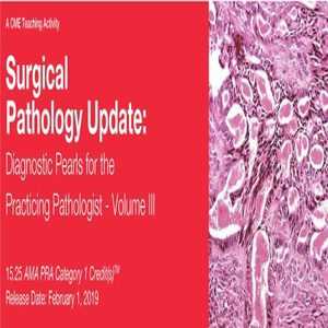 2019 Surgical Pathology Update Diagnostic Pearls for the Practicing Pathologist Vol. III (CME VIDEOS)
1 ×
2019 Surgical Pathology Update Diagnostic Pearls for the Practicing Pathologist Vol. III (CME VIDEOS)
1 ×30.00 $20.10 $ -
×
 Wall Breast and Body Contouring Video Library, Volume 2 QMP
1 ×
Wall Breast and Body Contouring Video Library, Volume 2 QMP
1 ×25.00 $16.75 $ -
×
 QMP Aesthetic Surgery of the Aging Face (6 DVD BOX)
1 ×
QMP Aesthetic Surgery of the Aging Face (6 DVD BOX)
1 ×50.00 $33.50 $ -
×
 Penn Radiology Comprehensive Neuroradiology: Optimal Care 2019
1 ×
Penn Radiology Comprehensive Neuroradiology: Optimal Care 2019
1 ×40.00 $26.80 $ -
×
 Cleveland Clinic Neurology Update OnDemand 2019
1 ×
Cleveland Clinic Neurology Update OnDemand 2019
1 ×30.00 $20.10 $ -
×
 Penn Medicine Penn Neuromuscular Symposium 2026
1 ×
Penn Medicine Penn Neuromuscular Symposium 2026
1 ×20.00 $13.40 $ -
×
 Cibas and Ducatman’s Cytology: Diagnostic Principles and Clinical Correlates, 6th edition (Original PDF from Publisher)
1 ×
Cibas and Ducatman’s Cytology: Diagnostic Principles and Clinical Correlates, 6th edition (Original PDF from Publisher)
1 ×30.00 $20.10 $ -
×
 ARRS A Synoptic Primer on the RADS 2021 (CME VIDEOS)
1 ×
ARRS A Synoptic Primer on the RADS 2021 (CME VIDEOS)
1 ×50.00 $33.50 $ -
×
 Gulfcoast Ultrasound Institute: Abdominal and Primary Care
1 ×
Gulfcoast Ultrasound Institute: Abdominal and Primary Care
1 ×35.00 $23.45 $ -
×
 UCSF : 55th Annual Recent Advances in Neurology 2022
1 ×
UCSF : 55th Annual Recent Advances in Neurology 2022
1 ×45.00 $30.15 $ -
×
 Harvard Comprehensive Review of Nephrology 2021 (CME Videos)
1 ×
Harvard Comprehensive Review of Nephrology 2021 (CME Videos)
1 ×10.00 $6.70 $ -
×
 Harvard Updates in General Medicine for Specialists (Videos + PDFs)
1 ×
Harvard Updates in General Medicine for Specialists (Videos + PDFs)
1 ×120.00 $80.40 $ -
×
 Oakstone The Brigham Update in Hospital Medicine 2026
1 ×
Oakstone The Brigham Update in Hospital Medicine 2026
1 ×38.00 $25.46 $ -
×
 ASPN Pediatric Nephrology Course 2021 Board Review And Refresher Course
1 ×
ASPN Pediatric Nephrology Course 2021 Board Review And Refresher Course
1 ×35.00 $23.45 $ -
×
 Echocardiography – A Comprehensive Review 2020
1 ×
Echocardiography – A Comprehensive Review 2020
1 ×40.00 $26.80 $ -
×
 USCAP How Senior Citizens from Cold Climates Deal with GI Biopsies in the Desert 2020 (An Emeritus Experience) 2020 (CME VIDEOS)
1 ×
USCAP How Senior Citizens from Cold Climates Deal with GI Biopsies in the Desert 2020 (An Emeritus Experience) 2020 (CME VIDEOS)
1 ×30.00 $20.10 $ -
×
 Topics in Mammography – 7th Edition 2019
1 ×
Topics in Mammography – 7th Edition 2019
1 ×30.00 $20.10 $ -
×
 SSRP Mastermind 2 Neurogenic Pain
2 ×
SSRP Mastermind 2 Neurogenic Pain
2 ×50.00 $33.50 $ -
×
 VIDEO Cosmetic Injection Techniques Neurotoxins and Fillers
1 ×
VIDEO Cosmetic Injection Techniques Neurotoxins and Fillers
1 ×25.00 $16.75 $ -
×
 Seeds Scientific Research & Performance Mastermind 14 The Brain Code 2025
1 ×
Seeds Scientific Research & Performance Mastermind 14 The Brain Code 2025
1 ×115.00 $77.05 $ -
×
 Penn Radiology – Breast Imaging State of the Art 2018
1 ×
Penn Radiology – Breast Imaging State of the Art 2018
1 ×15.00 $10.05 $ -
×
 Complete Dentures Simplified
1 ×
Complete Dentures Simplified
1 ×10.00 $6.70 $

OHI-S Microscope Ergonomics for Minimally Invasive Work Protocols
40.00 $
This Product is shared via google drive download link, So please share your correct Gmail id while placing the order .Please note that there are no CME points or certificate associated with this course
Samples for Courses Can be found here : Free Samples Here!
Description
Dive into minimally invasive techniques with tips and protocols on Microscopic Dentistry!
Video protocols from leading specialists Dr. Jenner Argueta and Cristian Coraini will guide you in successfully implementing a dental microscope into clinical prosthodontics, endodontics, and restorative dentistry.
During the course, you will learn in detail about:
– Protocols for setting up the dental microscope
– Ergonomics protocols for optimizing clinical workflow
– 3D microscope applications for minimally invasive direct and indirect restorations
– 3D microscope applications in endodontics and apical surgery.
Lesson 1.Introduction to 3D Microscopy in Dentistry
– Overview of 3d microscopy
– Transition from conventional dentistry to micro-dentistry
– Advantages of 3d microscopy: depth of field, imaging, ergonomics
– Introduction to 3d microscope features: magnification and illumination
– The learning curve and reducing frustration during operations with a 3D microscope.
Recommended for: Prosthodontists, Endodontists, Restorative dentist, General dentists.
Lesson 2.Ergonomics and Practical Tips for 3D Microscopy
– The importance of ergonomics in dentistry
– Patient positioning for effective use of 3D microscopy
– Protocols for reducing neck and back strain with 3D microscopy
– Tips for overcoming the learning curve
– Techniques for optimizing workflow and efficiency with 3D microscopy
– Documentation and teaching opportunities with 3D microscopy.
Recommended for: Prosthodontists, Endodontists, Restorative dentist, General dentists.
Lesson 3.Clinical Applications of 3D Microscopy
– The use of 3D microscopy in operative dentistry
– Application of 3D microscopy in endodontics
– Application of the 3D microscope in prosthodontics: crown and veneer preparation
– Application of the 3D microscope in microsurgery
– Protocols for enhancing clinical results with 3D imaging.
Recommended for: Prosthodontists, Endodontists, Restorative dentist, General dentists.
Lesson 4.Magnification in Dentistry: A Solution for Operational Difficulties
– Vision with the naked eye vs. Galilean and prismatic magnifying systems
– Reasons for using magnification in dentistry
– Operational difficulties for the operator with and without magnification
– Key differences between Galilean and prismatic systems, and operative microscopy
– The learning curve: practical tips and common errors to avoid.
Recommended for: Prosthodontists, Endodontists, Restorative dentist, General dentists.
Lesson 5.Setting up the Working Field: Ergonomics in Different Dental Disciplines
– Basic anatomy of the operating microscope (OM): optics, lenses, focal lengths, and lighting systems
– Practical tips for using surgical microscopy effectively
– Key settings: inter-pupillary distance, dioptric adjustment, and parfocality
– Magnification power, depth of field, and working distance
– Ergonomics in microscopy: operating positions and procedures in different dental disciplines.
Recommended for: Prosthodontists, Endodontists, Restorative dentist, General dentists.
Lesson 6.Minimally Invasive Dentistry (MID) with a Dental Microscope
– Clinical applications of magnification in minimally invasive prosthodontics and endodontics
– Minimally Invasive Dentistry (MID): significance and literature review
– Enhancing clinical results with microscopic magnification
– Intra-operative photos and videos for documentation and quality control
– How magnification improves precision, confidence, and treatment longevity.
Recommended for: Prosthodontists, Endodontists, Restorative dentist, General dentists.
Related products


